Current issue
About the Journal
Scientific Council
Editorial Board
Regulatory and archival policy
Code of publishing ethics
Publisher
Information about the processing of personal data in relation to cookies and newsletter subscription
Archive
For Authors
For Reviewers
Contact
Reviewers
Annals reviewers in 2025
Annals reviewers in 2024
Annals reviewers in 2023
Annals reviewers in 2022
Annals reviewers in 2021
Annals reviewers in 2020
Annals reviewers in 2019
Annals reviewers in 2018
Annals reviewers in 2017
Annals reviewers in 2016
Annals reviewers in 2015
Annals reviewers in 2014
Annals reviewers in 2013
Annals reviewers in 2012
Links
Sklep Wydawnictwa SUM
Biblioteka Główna SUM
Śląski Uniwersytet Medyczny w Katowicach
Privacy policy
Accessibility statement
Reviewers
Annals reviewers in 2025
Annals reviewers in 2024
Annals reviewers in 2023
Annals reviewers in 2022
Annals reviewers in 2021
Annals reviewers in 2020
Annals reviewers in 2019
Annals reviewers in 2018
Annals reviewers in 2017
Annals reviewers in 2016
Annals reviewers in 2015
Annals reviewers in 2014
Annals reviewers in 2013
Annals reviewers in 2012
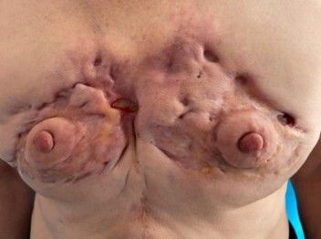
Morphea profunda – a case report of deep localized scleroderma with severe thorax deformity
1
Students’ Scientific Club, Department of Dermatology, Faculty of Medical Sciences in Katowice, Medical University of Silesia, Katowice, Poland
2
Department of Dermatology, Faculty of Medical Sciences in Katowice, Medical University of Silesia, Katowice, Poland
Corresponding author
Natalia Tekiela
Studenckie Koło Naukowe, Katedra i Klinika Dermatologii, ul. Francuska 20/24, SPSK im. A. Mielęckiego ŚUM, 40-027 Katowice
Studenckie Koło Naukowe, Katedra i Klinika Dermatologii, ul. Francuska 20/24, SPSK im. A. Mielęckiego ŚUM, 40-027 Katowice
Ann. Acad. Med. Siles. 2025;79:8-11
KEYWORDS
TOPICS
ABSTRACT
Deep scleroderma is a rare form of limited scleroderma characterised by deep sclerosis that may involve the muscles, fascia, subcutaneous tissue and deep layers of the skin. The lesions usually occur in the paraspinal line and may be predisposed by factors such as infections, injuries, exposure to radiation or the use of stimulants. Due to an insufficient level of awareness among healthcare professionals, diagnosis can be significantly delayed, and irreversible as well as crippling deformities can result. The study presents the case of a 67-year-old female patient with deep scleroderma whose lesions occur in an unusual location on the anterior surface of the thorax. This case demonstrates the importance of early diagnosis and the introduction of appropriate therapy in the active phase of the disease to avoid such severe consequences.
REFERENCES (11)
1.
Wolska-Gawron K., Krasowska D. Localized scleroderma – classification and tools used for the evaluation of tissue damage and disease activity/severity. Dermatol. Rev./Przegl. Dermatol. 2017; 104(3): 269–289, doi: 10.5114/dr.2017.68775.
2.
Cassisa A., Vannucchi M. Morphea profunda with tertiary lymphoid follicles: Description of two cases and review of the literature. Dermatopathology (Basel) 2022; 9(1): 17–22, doi: 10.3390/dermatopathology9010003.
3.
Shajil C., Sathishkumar D., Babu J.S., Babu R., Kumar S. Spontaneous crateriform indentation in a child. Pediatr. Dermatol. 2023; 40(4): 715–717, doi: 10.1111/pde.15241.
4.
Krasowska D., Rudnicka L., Dańczak-Pazdrowska A., Chodorowska G., Woźniacka A., Lis-Święty A. et al. Localized scleroderma (morphea): Diagnostic and therapeutic recommendations of the Polish Dermatological Society. Dermatol. Rev./Przegl. Dermatol. 2019; 106(4): 333–353, doi: 10.5114/dr.2019.88252.
5.
Budamakuntla L., Malvankar D. Extensive morphea profunda with autoantibodies and benign tumors: A rare case report. Indian Dermatol. Online J. 2012; 3(3): 208–210, doi: 10.4103/2229-5178.101823.
6.
Anggawirya B.Y., Indramaya D.M., Wardhani P.H., Widia Y., Citrashanty I., Sawitri S. et al. Deep circumscribed morphea: A case report. J. Pak. Assoc. Dermatol. 2024; 34(2): 582–586.
7.
Horger M., Fierlbeck G., Kuemmerle-Deschner J., Tzaribachev N., Wehrmann M., Claussen C.D. et al. MRI findings in deep and generalized morphea (localized scleroderma). AJR Am. J. Roentgenol. 2008; 190(1): 32–39, doi: 10.2214/AJR.07.2163.
8.
Khorasanizadeh F., Kalantari Y., Etesami I. Role of imaging in morphea assessment: A review of the literature. Skin Res. Technol. 2023; 29(7): e13410, doi: 10.1111/srt.13410.
9.
Penmetsa G.K., Sapra A. Morphea. StatPearls, 2023 [online]. Treasure Island (FL): StatPearls Publishing; 2024, https://www.ncbi.nlm.nih.gov/b... [accessed on 27 November 2024].
10.
Wolska-Gawron K., Michalska-Jakubus M., Krasowska D. Localized scleroderma – current treatment options. Dermatol. Rev./Przegl. Dermatol. 2017; 104(6): 606–618, doi: 10.5114/dr.2017.71833.
11.
Florez-Pollack S., Kunzler E., Jacobe H.T. Morphea: Current concepts. Clin. Dermatol. 2018; 36(4): 475–486, doi: 10.1016/j.clindermatol.2018.04.005.
The Medical University of Silesia in Katowice, as the Operator of the annales.sum.edu.pl website, processes personal data collected when visiting the website. The function of obtaining information about Users and their behavior is carried out by voluntarily entered information in forms, saving cookies in end devices, as well as by collecting web server logs, which are in the possession of the website Operator. Data, including cookies, are used to provide services in accordance with the Privacy policy.
You can consent to the processing of data for these purposes, refuse consent or access more detailed information.
You can consent to the processing of data for these purposes, refuse consent or access more detailed information.


