Current issue
Accepted manuscript
About the Journal
Scientific Council
Editorial Board
Regulatory and archival policy
Code of publishing ethics
Publisher
Information about the processing of personal data in relation to cookies and newsletter subscription
Archive
For Authors
For Reviewers
Contact
Reviewers
Annals reviewers in 2025
Annals reviewers in 2024
Annals reviewers in 2023
Annals reviewers in 2022
Annals reviewers in 2021
Annals reviewers in 2020
Annals reviewers in 2019
Annals reviewers in 2018
Annals reviewers in 2017
Annals reviewers in 2016
Annals reviewers in 2015
Annals reviewers in 2014
Annals reviewers in 2013
Annals reviewers in 2012
Links
Sklep Wydawnictwa SUM
Biblioteka Główna SUM
Śląski Uniwersytet Medyczny w Katowicach
Privacy policy
Accessibility statement
Reviewers
Annals reviewers in 2025
Annals reviewers in 2024
Annals reviewers in 2023
Annals reviewers in 2022
Annals reviewers in 2021
Annals reviewers in 2020
Annals reviewers in 2019
Annals reviewers in 2018
Annals reviewers in 2017
Annals reviewers in 2016
Annals reviewers in 2015
Annals reviewers in 2014
Annals reviewers in 2013
Annals reviewers in 2012

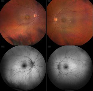
Presumed Doyne honeycomb macular dystrophy in two 72-year old twin sisters – case report
1
Students’ Scientific Club, Department of Ophthalmology, Faculty of Medical Sciences in Katowice, Medical University of Silesia, Katowice, Poland
2
Department of Ophthalmology, Faculty of Medical Sciences in Katowice, Medical University of Silesia, Katowice, Poland
These authors had equal contribution to this work
Corresponding author
Mikołaj Guzikowski
Studenckie Koło Naukowe, Klinika Okulistyki, Wydział Nauk Medycznych ŚUM, ul. Ceglana 35, 40-514 Katowice
Studenckie Koło Naukowe, Klinika Okulistyki, Wydział Nauk Medycznych ŚUM, ul. Ceglana 35, 40-514 Katowice

KEYWORDS
TOPICS
ABSTRACT
Introduction:
Hereditary macular dystrophies constitute a group of rare disorders whose exact prevalence is unknown. They include Doyne honeycomb retinal dystrophy (DHRD), inherited in an autosomal dominant manner and associated with a missense mutation in the EFEMP1 gene (2p16.1), which encodes a fibulin-like extracellular matrix protein containing an epidermal growth factor. Radially arranged, laminar, round drusen appear in childhood, whereas visual disturbances manifest only in the 4th and 5th decades of life.
Case reports:
Two 72-year old female patients, who are twins, presented to the Outpatient Ophthalmology Clinic due to visual disturbances. Visual acuity (VA) was 0.04 in the right eye (RE) and 0.08 in the left eye (LE) in patient A, and 0.06 in RE and 0.2 in LE in patient B. Intraocular pressure (IOP) measured 18 mmHg in RE and 17 mmHg in LE in patient A, and 17 mmHg in RE and 11 mmHg in LE in patient B. The patients were qualified for further diagnostic evaluation of both eyes.
Results:
OCT examination revealed confluent drusen and atrophy of the retinal pigment epithelium and photoreceptors in both patients. In patient A, an atrophic macular hole was additionally identified. The ganglion cell complex (GCC) measured an average of 120 μm in the RE and 120 μm in the LE in patient A, and 112 μm in RE and 113 μm in LE in patient B.
Conclusions:
Patients with suspected DHRD require regular ophthalmological follow-up to monitor disease progression, correct refractive errors, and assess the degree of lens opacity.
Hereditary macular dystrophies constitute a group of rare disorders whose exact prevalence is unknown. They include Doyne honeycomb retinal dystrophy (DHRD), inherited in an autosomal dominant manner and associated with a missense mutation in the EFEMP1 gene (2p16.1), which encodes a fibulin-like extracellular matrix protein containing an epidermal growth factor. Radially arranged, laminar, round drusen appear in childhood, whereas visual disturbances manifest only in the 4th and 5th decades of life.
Case reports:
Two 72-year old female patients, who are twins, presented to the Outpatient Ophthalmology Clinic due to visual disturbances. Visual acuity (VA) was 0.04 in the right eye (RE) and 0.08 in the left eye (LE) in patient A, and 0.06 in RE and 0.2 in LE in patient B. Intraocular pressure (IOP) measured 18 mmHg in RE and 17 mmHg in LE in patient A, and 17 mmHg in RE and 11 mmHg in LE in patient B. The patients were qualified for further diagnostic evaluation of both eyes.
Results:
OCT examination revealed confluent drusen and atrophy of the retinal pigment epithelium and photoreceptors in both patients. In patient A, an atrophic macular hole was additionally identified. The ganglion cell complex (GCC) measured an average of 120 μm in the RE and 120 μm in the LE in patient A, and 112 μm in RE and 113 μm in LE in patient B.
Conclusions:
Patients with suspected DHRD require regular ophthalmological follow-up to monitor disease progression, correct refractive errors, and assess the degree of lens opacity.
CONFLICT OF INTEREST
All the authors declare that they have no conflicts of interest.
Use of AI tools statement: ChatGPT was used for language translation of the manuscript.
REFERENCES (22)
1.
Chawla H, Tripathy K, Vohra V. Retinal Dystrophies. [Updated 2024 Oct 29]. In: StatPearls [Internet]. Treasure Island (FL): StatPearls Publishing; 2024 Jan-.
2.
Cusumano A, Falsini B, D’Ambrosio M, D’Apolito F, Sebastiani J, Levialdi Ghiron JH, et al. Long-Term Structural and Functional Assessment of Doyne Honeycomb Retinal Dystrophy following Nanosecond 2RT Laser Treatment: A Case Series. Case Rep Ophthalmol. 2023;14(1):626–639. doi: 10.1159/000534579.
3.
Zhang T, Xie X, Cao G, Jiang H, Wu S, Su Z, et al. Malattia leventinese/Doyne honeycomb retinal dystrophy in a Chinese family with mutation of the EFEMP1 gene. Retina. 2014;34(12):2462–2471. doi: 10.1097/IAE.0000000000000259.
4.
Tsang SH, Sharma T. Doyne Honeycomb Retinal Dystrophy (Malattia Leventinese, Autosomal Dominant Drusen). Adv Exp Med Biol. 2018;1085:97–102. doi: 10.1007/978-3-319-95046-4_18.
5.
Parameswarappa DC, Rani PK. Utility of pattern recognition and multimodal imaging in the diagnosis and management of doyne honeycomb retinal dystrophy complicated with type one choroidal neovascular membrane. BMJ Case Rep. 2021;14(2):e237635. doi: 10.1136/bcr-2020-237635.
6.
Shaw EM, Tate AJ, Periasamy R, Lipinski DM. Characterization of drusen formation in a primary porcine tissue culture model of dry AMD. Mol Ther Methods Clin Dev. 2024;32(4):101331. doi: 10.1016/j.omtm.2024.101331.
7.
Kim KL, Joo K, Park SJ, Park KH, Woo SJ. Progression from intermediate to neovascular age-related macular degeneration according to drusen subtypes: Bundang AMD cohort study report 3. Acta Ophthalmol. 2022;100(3):e710–e718. doi: 10.1111/aos.14960.
8.
Tong Y, Ach T, Curcio CA, Smith RT. Hyperspectral autofluorescence characterization of drusen and sub-RPE deposits in age-related macular degeneration. Ann Eye Sci. 2021;6:4. doi: 10.21037/aes-20-12.
9.
Curcio CA. Soft Drusen in Age-Related Macular Degeneration: Biology and Targeting Via the Oil Spill Strategies. Invest Ophthalmol Vis Sci. 2018;59(4):AMD160-AMD181. doi: 10.1167/iovs.18-24882.
10.
Curcio CA, Johnson M, Rudolf M, Huang JD. The oil spill in ageing Bruch membrane. Br J Ophthalmol. 2011;95(12):1638–1645. doi: 10.1136/bjophthalmol-2011-300344.
11.
Hulleman JD. Malattia Leventinese/Doyne Honeycomb Retinal Dystrophy: Similarities to Age-Related Macular Degeneration and Potential Therapies. Adv Exp Med Biol. 2016;854:153–158. doi: 10.1007/978-3-319-17121-0_21.
12.
Sohn EH, Wang K, Thompson S, Riker MJ, Hoffmann JM, Stone EM, et al. Comparison of drusen and modifying genes in autosomal dominant radial drusen and age-related macular degeneration. Retina. 2015;35(1):48–57. doi: 10.1097/IAE.0000000000000263.
13.
Querques G, Guigui B, Leveziel N, Querques L, Bandello F, Souied EH. Multimodal morphological and functional characterization of Malattia Leventinese. Graefes Arch Clin Exp Ophthalmol. 2013;251(3):705–714. doi: 10.1007/s00417-012-2106-5.
14.
AREDS2 Research Group; Chew EY, Clemons T, SanGiovanni JP, Danis R, Domalpally A, McBee W, et al. The Age-Related Eye Disease Study 2 (AREDS2): study design and baseline characteristics (AREDS2 report number 1). Ophthalmology. 2012;119(11):2282–2289. doi: 10.1016/j.ophtha.2012.05.027.
15.
Algvere PV, Kvanta A, Seregard S. Drusen maculopathy: a risk factor for visual deterioration. Acta Ophthalmol. 2016;94(5):427–433. doi: 10.1111/aos.13011.
16.
Misiuk-Hojło M, Bakunowicz-Łazarczyk A, Dobrowolski D, Grabska-Liberek I, Mackiewicz J, Mrukwa-Kominek E et al. Position Statement of the Polish Society of Ophthalmology establishing standards for the management of patients with exudative age-related macular degeneration. Klin Oczna. 2023;125(4):181–189. doi: 10.5114/ko.2023.134018.
17.
Professional Association of German Ophthalmologists (Berufsverband der Augenärzte Deutschlands e. V., BVA); German Society of Ophthalmology (Deutsche Ophthalmologische Gesellschaft, DOG); German Retina Society (Retinologische Gesellschaft e. V., RG). Statement and supplementary statement from the BVA, the DOG, and the RG on laser treatment of drusen in age-related macular degeneration (AMD) : August 2017, update October 2018. Ophthalmologe. 2020;117(Suppl 1):1–10. doi: 10.1007/s00347-019-0889-z.
18.
Kim KL, Han JM, Kim MS, Park SJ, Kim SW, Kim JH, et al. MACULAR HOLE ASSOCIATED WITH AGE-RELATED MACULAR DEGENERATION: Pathogenesis and Surgical Outcomes. Retina. 2021;41(10):2079–2087. doi: 10.1097/IAE.0000000000003148.
19.
Michalewska Z, Nawrocki J. Vitrectomy with the inverted internal limiting membrane flap technique in eyes with full-thickness macular hole and dry age-related macular degeneration. Eur J Ophthalmol. 2021;31(3):1320–1325. doi: 10.1177/1120672120921376.
20.
Tarkova A, Hejsek L, Jurecka T, Wolkova P, Rubesova M, Hackl M. Pars plana vitrectomy for vitreoretinal interface disorders coincident with intermediate stage age-related macular degeneration. Biomed Pap Med Fac Univ Palacky Olomouc Czech Repub. 2023;167(4):366–369. doi: 10.5507/bp.2022.047.
21.
Berzack S, Parekh PK. Drusen Regression Following Macular Hole Surgery: A Case Report. Retin Cases Brief Rep. 2026;20(2):282–284. doi: 10.1097/ICB.0000000000001720.
22.
Berinstein DM, Hassan TS, Williams GA, Margherio RR, Ruby AJ, Garretson BR. Surgical repair of full-thickness idiopathic macular holes associated with significant macular drusen. Ophthalmology. 2000;107(12):2233–2239. doi: 10.1016/s0161-6420(00)00417-6.
| eISSN: | 1734-025X |


The Medical University of Silesia in Katowice, as the Operator of the annales.sum.edu.pl website, processes personal data collected when visiting the website. The function of obtaining information about Users and their behavior is carried out by voluntarily entered information in forms, saving cookies in end devices, as well as by collecting web server logs, which are in the possession of the website Operator. Data, including cookies, are used to provide services in accordance with the Privacy policy.
You can consent to the processing of data for these purposes, refuse consent or access more detailed information.
You can consent to the processing of data for these purposes, refuse consent or access more detailed information.