Current issue
Accepted manuscript
About the Journal
Scientific Council
Editorial Board
Regulatory and archival policy
Code of publishing ethics
Publisher
Information about the processing of personal data in relation to cookies and newsletter subscription
Archive
For Authors
For Reviewers
Contact
Reviewers
Annals reviewers in 2025
Annals reviewers in 2024
Annals reviewers in 2023
Annals reviewers in 2022
Annals reviewers in 2021
Annals reviewers in 2020
Annals reviewers in 2019
Annals reviewers in 2018
Annals reviewers in 2017
Annals reviewers in 2016
Annals reviewers in 2015
Annals reviewers in 2014
Annals reviewers in 2013
Annals reviewers in 2012
Links
Sklep Wydawnictwa SUM
Biblioteka Główna SUM
Śląski Uniwersytet Medyczny w Katowicach
Privacy policy
Accessibility statement
Reviewers
Annals reviewers in 2025
Annals reviewers in 2024
Annals reviewers in 2023
Annals reviewers in 2022
Annals reviewers in 2021
Annals reviewers in 2020
Annals reviewers in 2019
Annals reviewers in 2018
Annals reviewers in 2017
Annals reviewers in 2016
Annals reviewers in 2015
Annals reviewers in 2014
Annals reviewers in 2013
Annals reviewers in 2012

From thread to yarn, and yarn to thread: a complex case of persistent left superior vena cava
1
1st Department of Cardiology, Faculty of Medical Sciences in Katowice, Medical University of Silesia, Katowice, Poland; Centre of the European Reference Network for Rare, Low Prevalence or Complex Diseases of the Heart (ERN GUARD Heart)
Corresponding author
Julia Dołęga
Klinika Kardiologii I Katedry Kardiologii, Śląski Uniwersytet Medyczny w Katowicach, ul. Ziołowa 47, 40-635 Katowice
Klinika Kardiologii I Katedry Kardiologii, Śląski Uniwersytet Medyczny w Katowicach, ul. Ziołowa 47, 40-635 Katowice
Ann. Acad. Med. Siles. 2025;79:24-28

KEYWORDS
TOPICS
ABSTRACT
Introduction:
Persistent left superior vena cava (PLSVC) is a rare venous anomaly, occurring in 0.3–0.5% of the general population and up to 4.3% of patients with heart defects. It forms from the junction of the left subclavian and internal jugular veins, passes through the left mediastinum, and drains into the right atrium via the coronary sinus. Usually asymptomatic, it is typically discovered incidentally during imaging and may be associated with an atrial septal defect (ASD).
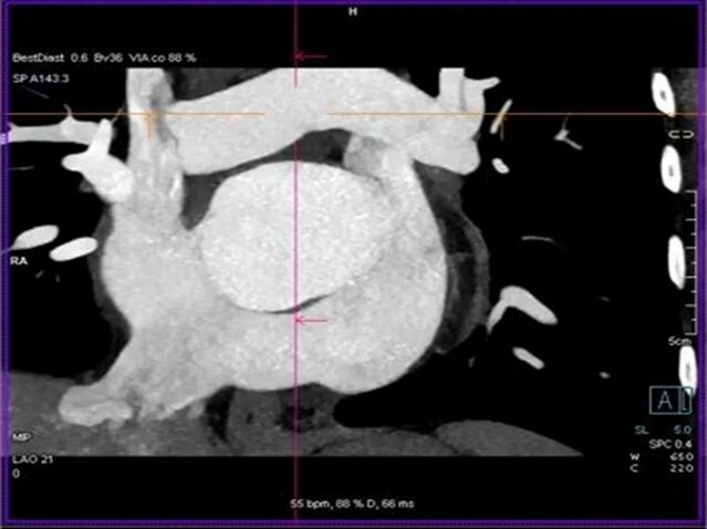
Case report:
A 52-year-old female patient with persistent atrial fibrillation, a history of ischemic stroke in the left hemisphere of the brain, uncontrolled hypertension, and diagnosed with ASD type 2, was referred for pulmonary vein isolation (PVI) due to symptomatic arrhythmia of European Heart Rhythm Association class IIb and New York Heart Association class II severity. After unsuccessful PVI, pharmacological cardioversion was attempted, followed by electrical cardioversion, which temporarily restored sinus rhythm. Echocardiography revealed moderate tricuspid valve regurgitation and an enlarged coronary sinus. Cardiac computed tomography was ordered, revealing the presence of a PLSVC, into which the left superior pulmonary vein drains, with rightward displacement of the interatrial septum and a patent foramen ovale (PFO). After cardiac surgery consultation, the patient was qualified for defect correction.
Conclusions:
PLSVC may be associated with congenital defects such as ASD type 2/PFO, which is relevant in the treatment of arrhythmias and defect correction. An enlarged coronary sinus on echocardiography should raise suspicion of PLSVC. The presence of PLSVC is significant when placing devices with central venous access.
Persistent left superior vena cava (PLSVC) is a rare venous anomaly, occurring in 0.3–0.5% of the general population and up to 4.3% of patients with heart defects. It forms from the junction of the left subclavian and internal jugular veins, passes through the left mediastinum, and drains into the right atrium via the coronary sinus. Usually asymptomatic, it is typically discovered incidentally during imaging and may be associated with an atrial septal defect (ASD).
Case report:
A 52-year-old female patient with persistent atrial fibrillation, a history of ischemic stroke in the left hemisphere of the brain, uncontrolled hypertension, and diagnosed with ASD type 2, was referred for pulmonary vein isolation (PVI) due to symptomatic arrhythmia of European Heart Rhythm Association class IIb and New York Heart Association class II severity. After unsuccessful PVI, pharmacological cardioversion was attempted, followed by electrical cardioversion, which temporarily restored sinus rhythm. Echocardiography revealed moderate tricuspid valve regurgitation and an enlarged coronary sinus. Cardiac computed tomography was ordered, revealing the presence of a PLSVC, into which the left superior pulmonary vein drains, with rightward displacement of the interatrial septum and a patent foramen ovale (PFO). After cardiac surgery consultation, the patient was qualified for defect correction.
Conclusions:
PLSVC may be associated with congenital defects such as ASD type 2/PFO, which is relevant in the treatment of arrhythmias and defect correction. An enlarged coronary sinus on echocardiography should raise suspicion of PLSVC. The presence of PLSVC is significant when placing devices with central venous access.
REFERENCES (8)
1.
Uemura T., Kondo H., Shinohara T., Takahashi M., Akamine K., Ogawa N. et al. Multiple accessory pathways coexisting with a persistent left superior vena cava: a case report. J. Med. Case Rep. 2023; 17(1): 111, doi: 10.1186/s13256-023-03865-6.
2.
Polewczyk A., Kutarski A., Czekajska-Chehab E., Adamczyk P., Boczar K., Polewczyk M. et al. Complications of permanent cardiac pacing in patients with persistent left superior vena cava. Cardiol. J. 2014; 21(2): 128–137, doi: 10.5603/CJ.a2014.0006.
3.
Batouty N.M., Sobh D.M., Gadelhak B., Sobh H.M., Mahmoud W., Tawfik A.M. Left superior vena cava: cross-sectional imaging overview. Radiol. Med. 2020; 125(3): 237–246, doi: 10.1007/s11547-019-01114-9.
4.
Higgs A.G., Paris S., Potter F. Discovery of left-sided superior vena cava during central venous catheterization. Br. J. Anaesth. 1998; 81(2): 260–261, doi: 10.1093/bja/81.2.260.
5.
Sarodia B.D., Stoller J.K. Persistent left superior vena cava: case report and literature review. Respir. Care 2000; 45(4): 411–416.
6.
Sonavane S.K., Milner D.M., Singh S.P., Abdel Aal A.K., Shahir K.S., Chaturvedi A. Comprehensive imaging review of the superior vena cava. Radiographics 2015; 35(7): 1873–1892, doi: 10.1148/rg.2015150056.
7.
Demos T.C., Posniak H.V., Pierce K.L., Olson M.C., Muscato M. Venous anomalies of the thorax. AJR Am. J. Roentgenol. 2004; 182(5): 1139–1150, doi: 10.2214/ajr.182.5.1821139.
8.
Stewart J.A., Fraker T.D. Jr, Slosky D.A., Wise N.K., Kisslo J.A. Detection of persistent left superior vena cava by two-dimensional contrast echocardiography. J. Clin. Ultrasound 1979; 7(5): 357–360, doi: 10.1002/jcu.1870070506.
CITATIONS (1):
1.
Persistent left superior vena cava as a challenge in atrial fibrillation ablation – successful isolation using PulseSelect
Aleksandra Kłosińska, Piotr Anders, Paweł Jesionowski
In a Good Rhythm
Aleksandra Kłosińska, Piotr Anders, Paweł Jesionowski
In a Good Rhythm
| eISSN: | 1734-025X |

The Medical University of Silesia in Katowice, as the Operator of the annales.sum.edu.pl website, processes personal data collected when visiting the website. The function of obtaining information about Users and their behavior is carried out by voluntarily entered information in forms, saving cookies in end devices, as well as by collecting web server logs, which are in the possession of the website Operator. Data, including cookies, are used to provide services in accordance with the Privacy policy.
You can consent to the processing of data for these purposes, refuse consent or access more detailed information.
You can consent to the processing of data for these purposes, refuse consent or access more detailed information.