Current issue
About the Journal
Scientific Council
Editorial Board
Regulatory and archival policy
Code of publishing ethics
Publisher
Information about the processing of personal data in relation to cookies and newsletter subscription
Archive
For Authors
For Reviewers
Contact
Reviewers
Annals reviewers in 2025
Annals reviewers in 2024
Annals reviewers in 2023
Annals reviewers in 2022
Annals reviewers in 2021
Annals reviewers in 2020
Annals reviewers in 2019
Annals reviewers in 2018
Annals reviewers in 2017
Annals reviewers in 2016
Annals reviewers in 2015
Annals reviewers in 2014
Annals reviewers in 2013
Annals reviewers in 2012
Links
Sklep Wydawnictwa SUM
Biblioteka Główna SUM
Śląski Uniwersytet Medyczny w Katowicach
Privacy policy
Accessibility statement
Reviewers
Annals reviewers in 2025
Annals reviewers in 2024
Annals reviewers in 2023
Annals reviewers in 2022
Annals reviewers in 2021
Annals reviewers in 2020
Annals reviewers in 2019
Annals reviewers in 2018
Annals reviewers in 2017
Annals reviewers in 2016
Annals reviewers in 2015
Annals reviewers in 2014
Annals reviewers in 2013
Annals reviewers in 2012
Detection of liver metastases by abbreviated MRI protocol in patient with pNET and severe chronic kidney disease
1
Department of Radiology and Nuclear Medicine, Medical University of Silesia, Katowice, Poland
Corresponding author
Mateusz Winder
Zakład Radiodiagnostyki i Radiologii Zabiegowej, Uniwersyteckie Centrum Kliniczne im. prof. K. Gibińskiego Śląskiego Uniwersytetu Medycznego w Katowicach, ul. Medyków 14, 40-752 Katowice
Zakład Radiodiagnostyki i Radiologii Zabiegowej, Uniwersyteckie Centrum Kliniczne im. prof. K. Gibińskiego Śląskiego Uniwersytetu Medycznego w Katowicach, ul. Medyków 14, 40-752 Katowice
Ann. Acad. Med. Siles. 2024;78:294-297
KEYWORDS
TOPICS
ABSTRACT
The detection of liver metastases at an early stage is crucial to improve patients’ survival. Pancreatic neuroendocrine tumors (pNETs) and their metastases usually present as early and vividly enhancing tumors on contrast enhanced
computed tomography (CECT). However, the use of contrast agents is a contraindication in patients with severely impaired kidney function. The incidence of chronic kidney disease (CKD) and malignancies increase with age, making both the treatment and imaging diagnostics a complicated process in an oncological setting among elderly patients.
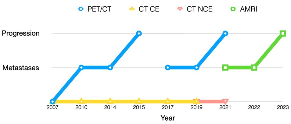
This case report presents the diagnostic path of an elderly patient with a long history of pNET, who developed severe CKD during treatment. The patient was diagnosed with metastatic liver disease in PET/CT and it was confirmed by an abbreviated magnetic resonance imaging (AMRI) protocol without contrast enhancement, while CECT did not show the presence of metastases. AMRI protocols without contrast enhancement can provide sufficient information about the presence of metastatic liver disease in oncological patients with comorbid CKD.
FUNDING
This research received no funding.
CONFLICT OF INTEREST
None declared.
REFERENCES (8)
1.
Mao Y., Chen B., Wang H., Zhang Y., Yi X., Liao W. et al. Diagnostic performance of magnetic resonance imaging for colorectal liver metastasis: A systematic review and meta-analysis. Sci. Rep. 2020; 10(1): 1969, doi: 10.1038/s41598-020-58855-1.
2.
Shaikh S. Editorial for “Abbreviated gadoxetic acid-enhanced MRI for the detection of liver metastases in patients with potentially resectable pancreatic ductal adenocarcinoma”. J. Magn. Reson. Imaging 2022; 56(3): 737–738, doi: 10.1002/jmri.28054.
3.
Granata C., Bicchierai G., Fusco R., Cozzi D., Grazzini G., Danti G. et al. Diagnostic protocols in oncology: workup and treatment planning. Part 2: Abbreviated MR protocol. Eur. Rev. Med. Pharmacol. Sci. 2021; 25(21): 6499–6528, doi: 10.26355/eurrev_202111_27094.
4.
Scott A.T., Howe J.R. Evaluation and management of neuroendocrine tumors of the pancreas. Surg. Clin. North Am. 2019; 99(4): 793–814, doi: 10.1016/j.suc.2019.04.014.
5.
Chiti G., Grazzini G., Cozzi D., Danti G., Matteuzzi B., Granata V. et al. Imaging of pancreatic neuroendocrine neoplasms. Int. J. Environ. Res. Public Health 2021; 18(17): 8895, doi: 10.3390/ijerph18178895.
6.
Winder M., Grabowska S., Hitnarowicz A., Barczyk-Gutkowska A., Gruszczyńska K., Steinhof-Radwańska K. The application of abbreviated MRI protocols in malignant liver lesions surveillance. Eur. J. Radiol. 2023; 164: 110840, doi: 10.1016/j.ejrad.2023.110840.
7.
Grabowska S., Hitnarowicz A., Barczyk-Gutkowska A., Gruszczyńska K., Steinhof-Radwańska K., Winder M. Abbreviated magnetic resonance imaging protocols in oncology: improving accessibility in precise diagnostics. Pol. J. Radiol. 2023; 88: e415–e422, doi: 10.5114/pjr.2023.131213.
8.
Bednarek A., Mykała-Cieśla J., Pogoda K., Jagiełło-Gruszfeld A., Kunkiel M., Winder M. et al. Limitations of systemic oncological therapy in breast cancer patients with chronic kidney disease. J. Oncol. 2020; 2020: 7267083, doi: 10.1155/2020/7267083.
Share
RELATED ARTICLE
The Medical University of Silesia in Katowice, as the Operator of the annales.sum.edu.pl website, processes personal data collected when visiting the website. The function of obtaining information about Users and their behavior is carried out by voluntarily entered information in forms, saving cookies in end devices, as well as by collecting web server logs, which are in the possession of the website Operator. Data, including cookies, are used to provide services in accordance with the Privacy policy.
You can consent to the processing of data for these purposes, refuse consent or access more detailed information.
You can consent to the processing of data for these purposes, refuse consent or access more detailed information.


