Bieżący numer
Artykuły zaakceptowane
O czasopiśmie
Rada Naukowa
Kolegium Redakcyjne
Polityka prawno-archiwizacyjna
Kodeks etyki publikacyjnej
Wydawca
Informacja o przetwarzaniu danych osobowych w ramach plików cookies oraz subskrypcji newslettera
Archiwum
Dla autorów
Dla recenzentów
Kontakt
Recenzenci
Recenzenci rocznika 2025
Recenzenci rocznika 2024
Recenzenci rocznika 2023
Recenzenci rocznika 2022
Recenzenci rocznika 2021
Recenzenci rocznika 2020
Recenzenci rocznika 2019
Recenzenci rocznika 2018
Recenzenci rocznika 2017
Recenzenci rocznika 2016
Recenzenci rocznika 2015
Recenzenci rocznika 2014
Recenzenci rocznika 2013
Recenzenci rocznika 2012
Polecamy
Śląski Uniwersytet Medyczny w Katowicach
Sklep Wydawnictw SUM
Biblioteka Główna SUM
Polityka prywatności
Deklaracja dostępności
Recenzenci
Recenzenci rocznika 2025
Recenzenci rocznika 2024
Recenzenci rocznika 2023
Recenzenci rocznika 2022
Recenzenci rocznika 2021
Recenzenci rocznika 2020
Recenzenci rocznika 2019
Recenzenci rocznika 2018
Recenzenci rocznika 2017
Recenzenci rocznika 2016
Recenzenci rocznika 2015
Recenzenci rocznika 2014
Recenzenci rocznika 2013
Recenzenci rocznika 2012

Zrozumienie trakcji szklistkowo-plamkowej: podstawowe pojęcia i znaczenie kliniczne
1
Students’ Scientific Club, Department of Ophthalmology, Faculty of Medical Sciences in Katowice, Medical University of Silesia, Katowice, Poland
2
Department of Ophthalmology, Faculty of Medical Sciences in Katowice, Medical University of Silesia, Katowice, Poland
Autor do korespondencji
Sabina Kowalczyk
Studenckie Koło Naukowe, Klinika Okulistyki, Wydział Nauk Medycznych ŚUM, ul. Ceglana 35, 40-514 Katowice
Studenckie Koło Naukowe, Klinika Okulistyki, Wydział Nauk Medycznych ŚUM, ul. Ceglana 35, 40-514 Katowice
Ann. Acad. Med. Siles. 2026;80:124-132

SŁOWA KLUCZOWE
zaburzenia interakcji szklistkowo-plamkowychtrakcja szklistkowo-plamkowaretinopatia cukrzycowawitrektomia pars plana
DZIEDZINY
STRESZCZENIE
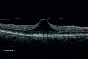
Pierwsze wzmianki o trakcji szklistkowo-plamkowej (vitreomacular traction – VMT) pochodzą z 1953 roku, gdy Samuel Rodman Irvine zidentyfikował ją jako potencjalną przyczynę torbielowatego obrzęku plamki. W ostatnich latach w literaturze coraz częściej pojawiają się doniesienia o powiązaniach między VMT a innymi powszechnymi schorzeniami oczu, takimi jak retinopatia cukrzycowa i zwyrodnienie plamki związane z wiekiem (age-related macular degeneration – AMD). Celem pracy jest omówienie epidemiologii, czynników ryzyka, patofizjologii, metod diagnostycznych oraz możliwości leczenia VMT. Mechanizm rozwoju VMT w przebiegu retinopatii cukrzycowej różni się w zależności od tego, czy mamy do czynienia z proliferacyjną czy nieproliferacyjną postacią retinopatii. Wysiękowa postać AMD charakteryzuje się rozwojem neowaskularyzacji plamkowej, prowadzącej do wysięku lub krwotoku w plamce. Najnowsze badania sugerują również możliwy związek między AMD i VMT. VMT oraz inne zaburzenia interakcji szklistkowo-siatkówkowych to grupa schorzeń o zróżnicowanych patomechanizmach i powikłaniach. Wybór skutecznej metody leczenia wymaga uwzględnienia wielu czynników klinicznych. Aby było to możliwe, lekarze zajmujący się tym zagadnieniem muszą posiadać solidną wiedzę. Niniejszy artykuł stanowi przegląd kluczowych zagadnień oraz aktualnego stanu wiedzy na temat VMT.
REFERENCJE (57)
1.
Irvine SR. A newly defined vitreous syndrome following cataract surgery. Am J Ophthalmol. 1953;36(5):599–619. doi: 10.1016/0002-9394(53)90302-x.
2.
Jaffe NS. Vitreous traction at the posterior pole of the fundus due to alterations in the vitreous posterior. Trans Am Acad Ophthalmol Otolaryngol. 1967;71(4):642–652.
3.
Reese AB, Jones IS, Cooper WC. Vitreomacular traction syndrome confirmed histologically. Am J Ophthalmol. 1970;69(6):975–977. doi: 10.1016/0002-9394(70)91041-x.
4.
Faatz H, Hattenbach LO, Krohne TU, Priglinger SG, Lommatzsch A. Vitreomacular traction: diagnostics, natural course, treatment decision and guideline recommendations. [Article in German]. Ophthalmologie. 2024;121(6):470–475. doi: 10.1007/s00347-024-02042-4.
5.
Duker JS, Kaiser PK, Binder S, de Smet MD, Gaudric A, Reichel E, et al. The International Vitreomacular Traction Study Group classification of vitreomacular adhesion, traction, and macular hole. Ophthalmology. 2013;120(12):2611–2619. doi: 10.1016/j.ophtha.2013.07.042.
6.
Menzler J, Neubauer AS, Haritoglou C, Jackson TL. Incidence and prevalence of vitreomacular traction with and without macular hole in Germany. Clin Ophthalmol. 2019;13:177–188. doi: 10.2147/OPTH.S188704.
7.
Ramovecchi P, Salati C, Zeppieri M. Spontaneous posterior vitreous detachment: A glance at the current literature. World J Exp Med. 2021;11(3):30–36. doi: 10.5493/wjem.v11.i3.30.
8.
Jones CH, Gui W, Schumann RG, Boneva S, Lange CA, van Overdam K, et al. Hyalocytes in proliferative vitreo-retinal diseases. Expert Rev Ophthalmol. 2022;17(4):263–280. doi: 10.1080/17469899.2022.2100764.
9.
Sebag J. Anomalous posterior vitreous detachment: a unifying concept in vitreo-retinal disease. Graefes Arch Clin Exp Ophthalmol. 2004;242(8):690–698. doi: 10.1007/s00417-004-0980-1.
10.
Rodman JA, Shechtman D, Sutton BM, Pizzimenti JJ, Bittner AK; VAST Study Group. Prevalence of vitreomacular adhesion in patients without maculopathy older than 40 years. Retina. 2018;38(10):2056–2063. doi: 10.1097/iae.0000000000001792.
11.
Bottós J, Elizalde J, Maia M. Natural history and pathogenesis of vitreomacular traction. Retina Today. 2014;(May–June):32–40.
12.
Punjabi OS, Kaiser PK. Vitreomacular Adhesion in Exudative Age-related Macular Degeneration. US Ophthalmic Rev. 2011;4(1):122–124. doi: 10.17925/USOR.2011.04.02.122.
13.
Simpson ARH, Petrarca R, Jackson TL. Vitreomacular Adhesion and Neovascular Age-Related Macular Degeneration. Surv Ophthalmol. 2012;57(6):498–509. doi: 10.1016/j.survophthal.2012.01.011.
14.
Xie P, Zheng X, Yu Y, Ye X, Hu Z, Yuan D, et al. Vitreomacular adhesion or vitreomacular traction may affect antivascular endothelium growth factor treatment for neovascular age-related macular degeneration. Br J Ophthalmol. 2017;101(8):1003–1010. doi: 10.1136/bjophthalmol-2017-310155.
15.
Lorusso M, Micelli Ferrari L, Leozappa M, Modoni AP, Micelli Ferrari T. Transient vitreomacular traction syndrome caused by traumatic incomplete posterior vitreous detachment. Eur J Ophthalmol. 2011;21(5):668–670. doi: 10.5301/EJO.2011.6425.
16.
Sebag J. Posterior Vitreous Detachment. Ophthalmology. 2018;125(9):1384–1385. doi: 10.1016/j.ophtha.2018.05.018.
17.
Wergenthaler N, Dick HB, Tsai T, Joachim SC. Etiology of Idiopathic Macular Holes in the Light of Estrogen Hormone. Curr Issues Mol Biol. 2023;45(8):6339–6351. doi: 10.3390/cimb45080400.
18.
Hsieh YT, Yang CM. Vitreomacular traction in diabetic retinopathy. Jpn J Ophthalmol. 2024;68(1):12–18. doi: 10.1007/s10384-023-01034-2.
19.
Haines NR, Manoharan N, Olson JL, D’Alessandro A, Reisz JA. Metabolomics Analysis of Human Vitreous in Diabetic Retinopathy and Rhegmatogenous Retinal Detachment. J Proteome Res. 2018;17(7):2421–2427. doi: 10.1021/acs.jproteome.8b00169.
20.
Huang H. Pentose phosphate pathway and diabetic retinopathy. Invest Ophthalmol Vis Sci. 2023;64(8):1823.
21.
Lin A, Xia H, Zhang A, Liu X, Chen H. Vitreomacular Interface Disorders in Proliferative Diabetic Retinopathy: An Optical Coherence Tomography Study. J Clin Med. 2022;11(12):3266. doi: 10.3390/jcm11123266.
22.
Li X, Luo X, Lu X, Duan J, Xu G. Metabolomics study of diabetic retinopathy using gas chromatography-mass spectrometry: a comparison of stages and subtypes diagnosed by Western and Chinese medicine. Mol Biosyst. 2011;7(7):2228–2237. doi: 10.1039/c0mb00341g.
23.
Klaassen I, van Geest RJ, Kuiper EJ, van Noorden CJ, Schlingemann RO. The role of CTGF in diabetic retinopathy. Exp Eye Res. 2015;133:37–48. doi: 10.1016/j.exer.2014.10.016.
24.
Hayashi K, Sato T, Manabe SI, Hirata A, Yoshimura K. Posterior vitreous detachment in patients with diabetes mellitus. Jpn J Ophthalmol. 2020;64(2):187–195. doi: 10.1007/s10384-020-00720-9.
25.
Perais J, Agarwal R, Evans JR, Loveman E, Colquitt JL, Owens D, et al. Prognostic factors for the development and progression of proliferative diabetic retinopathy in people with diabetic retinopathy. Cochrane Database Syst Rev. 2023;2(2):CD013775. doi: 10.1002/14651858.CD013775.pub2.
26.
Rangasamy S, McGuire PG, Franco Nitta C, Monickaraj F, Oruganti SR, Das A. Chemokine mediated monocyte trafficking into the retina: role of inflammation in alteration of the blood-retinal barrier in diabetic retinopathy. PLoS One. 2014;9(10):e108508. doi: 10.1371/journal.pone.0108508.
27.
Su CC, Yang CH, Yeh PT, Yang CM. Macular tractional retinoschisis in proliferative diabetic retinopathy: clinical characteristics and surgical outcome. Ophthalmologica. 2014;231(1):23–30. doi: 10.1159/000355078.
28.
Iyer SSR, Lagrew MK, Tillit SM, Roohipourmoallai R, Korntner S. The Vitreous Ecosystem in Diabetic Retinopathy: Insight into the Patho-Mechanisms of Disease. Int J Mol Sci. 2021;22(13):7142. doi: 10.3390/ijms22137142.
29.
Agarwal D, Gelman R, Prospero Ponce C, Stevenson W, Christoforidis JB. The Vitreomacular Interface in Diabetic Retinopathy. J Ophthalmol. 2015;2015:392983. doi: 10.1155/2015/392983.
30.
Petrovič MG, Korošec P, Košnik M, Hawlina M. Association of preoperative vitreous IL-8 and VEGF levels with visual acuity after vitrectomy in proliferative diabetic retinopathy. Acta Ophthalmol. 2010;88(8):e311–316. doi: 10.1111/j.1755-3768.2010.02030.x.
31.
Fleckenstein M, Keenan TDL, Guymer RH, Chakravarthy U, Schmitz-Valckenberg S, Klaver CC, et al. Age-related macular degeneration. Nat Rev Dis Primers. 2021;7(1):31. doi: 10.1038/s41572-021-00265-2.
32.
Schulze S, Hoerle S, Mennel S, Kroll P. Vitreomacular traction and exudative age-related macular degeneration. Acta Ophthalmol. 2008;86(5):470–481. doi: 10.1111/j.1755-3768.2008.01210.x.
33.
Green-Simms AE, Bakri SJ. Vitreomacular traction and age-related macular degeneration. Semin Ophthalmol. 2011;26(3):137–138. doi: 10.3109/08820538.2011.559512.
34.
Chen M, Xu H. Parainflammation, chronic inflammation, and age-related macular degeneration. J Leukoc Biol. 2015;98(5):713–725. doi: 10.1189/jlb.3RI0615-239R.
35.
Kang EC, Koh HJ. Effects of Vitreomacular Adhesion on Age-Related Macular Degeneration. J Ophthalmol. 2015;2015:865083. doi: 10.1155/2015/865083.
36.
Ondeş F, Yilmaz G, Acar MA, Unlü N, Kocaoğlan H, Arsan AK. Role of the vitreous in age-related macular degeneration. Jpn J Ophthalmol. 2000;44(1):91–93. doi: 10.1016/s0021-5155(99)00174-4.
37.
Krebs I, Brannath W, Glittenberg C, Zeiler F, Sebag J, Binder S. Posterior vitreomacular adhesion: a potential risk factor for exudative age-related macular degeneration? Am J Ophthalmol. 2007;144(5):741–746. doi: 10.1016/j.ajo.2007.07.024.
38.
Do DV, Cho M, Nguyen QD, Shah SM, Handa JT, Campochiaro PA, et al. The impact of optical coherence tomography on surgical decision making in epiretinal membrane and vitreomacular traction. Trans Am Ophthalmol Soc. 2006;104:161–166.
39.
Steel DHW, Downey L, Greiner K, Heimann H, Jackson TL, Koshy Z, et al. The design and validation of an optical coherence tomography-based classification system for focal vitreomacular traction. Eye (Lond). 2016;30(2):314–324; quiz 325. doi: 10.1038/eye.2015.262.
40.
Gass JD. Reappraisal of biomicroscopic classification of stages of development of a macular hole. Am J Ophthalmol. 1995;119(6):752–759. doi: 10.1016/s0002-9394(14)72781-3.
41.
Bottós J, Elizalde J, Arevalo JF, Rodrigues EB, Maia M. Vitreomacular traction syndrome. J Ophthalmic Vis Res. 2012;7(2):148–161.
42.
Ketkar M, Dave VP, de Ribot FM, Sallam AB, Shettigar MP, Hsieh YT, et al. Vitreomacular traction – a review. Eye (Lond). 2025;39(4):710–717. doi: 10.1038/s41433-024-03576-2.
43.
Theodossiadis GP, Grigoropoulos VG, Theodoropoulou S, Datseris I, Theodossiadis PG. Spontaneous resolution of vitreomacular traction demonstrated by spectral-domain optical coherence tomography. Am J Ophthalmol. 2014;157(4):842–851.e1. doi: 10.1016/j.ajo.2014.01.011.
44.
Hikichi T, Yoshida A, Trempe CL. Course of vitreomacular traction syndrome. Am J Ophthalmol. 1995;119(1):55–61. doi: 10.1016/s0002-9394(14)73813-9.
45.
Errera MH, Liyanage SE, Petrou P, Keane PA, Moya R, Ezra E, et al. A Study of the Natural History of Vitreomacular Traction Syndrome by OCT. Ophthalmology. 2018;125(5):701–707. doi: 10.1016/j.ophtha.2017.10.035.
46.
Dimopoulos S, Bartz-Schmidt KU, Gelisken F, Januschowski K, Ziemssen F. Rate and timing of spontaneous resolution in a vitreomacular traction group: should the role of watchful waiting be re-evaluated as an alternative to ocriplasmin therapy? Br J Ophthalmol. 2015;99(3):350–353. doi: 10.1136/bjophthalmol-2014-304961.
47.
Nazari H, Modarres-Zadeh M, Maleki A. Pharmacologic vitreolysis. J Ophthalmic Vis Res. 2010;5(1):44–52.
48.
Prospero Ponce CM, Stevenson W, Gelman R, Agarwal DR, Christoforidis JB. Ocriplasmin: who is the best candidate? Clin Ophthalmol. 2016;10:485–495. doi: 10.2147/OPTH.S97947.
49.
Stalmans P, Benz MS, Gandorfer A, Kampik A, Girach A, Pakola S, et al. Enzymatic vitreolysis with ocriplasmin for vitreomacular traction and macular holes. N Engl J Med. 2012;367(7):606–615. doi: 10.1056/NEJMoa1110823.
50.
Bennison C, Stephens S, Lescrauwaet B, Van Hout B, Jackson TL. Cost-effectiveness of ocriplasmin for the treatment of vitreomacular traction and macular hole. J Mark Access Health Policy. 2016;4. doi: 10.3402/jmahp.v4.31472.
51.
Morescalchi F, Gambicorti E, Duse S, Costagliola C, Semeraro F. From the analysis of pharmacologic vitreolysis to the comprehension of ocriplasmin safety. Expert Opin Drug Saf. 2016;15(9):1267–1278. doi: 10.1080/14740338.2016.1208169.
52.
Johnson MW. How should we release vitreomacular traction: surgically, pharmacologically, or pneumatically? Am J Ophthalmol. 2013;155(2):203–205.e1. doi: 10.1016/j.ajo.2012.10.016.
53.
Rodrigues IA, Stangos AN, McHugh DA, Jackson TL. Intravitreal injection of expansile perfluoropropane (C3F8) for the treatment of vitreomacular traction. Am J Ophthalmol 2013; 155(2):270–276.e2. doi: 10.1016/j.ajo.2012.08.018.
54.
Chan CK, Mein CE, Glassman AR, Beaulieu WT, Calhoun CT, Jaffe GJ, et al. Pneumatic Vitreolysis with Perfluoropropane for Vitreomacular Traction with and without Macular Hole: DRCR Retina Network Protocols AG and AH. Ophthalmology. 2021;128(11):1592–1603. doi: 10.1016/j.ophtha.2021.05.005.
55.
Flynn HW Jr, Relhan N. The Charles Schepens Lecture: Management Options for Vitreomacular Traction: Use an Individualized Approach. Ophthalmol Retina. 2017;1(1):3–7. doi: 10.1016/j.oret.2016.09.006.
56.
García-Layana A, García-Arumí J, Ruiz-Moreno JM, Arias-Barquet L, Cabrera-López F, Figueroa MS. A review of current management of vitreomacular traction and macular hole. J Ophthalmol. 2015;2015:809640. doi: 10.1155/2015/809640.
57.
Quiroz-Reyes MA, Quiroz-Gonzalez EA, Quiroz-Gonzalez MA, Lima-Gomez V. Pneumatic vitreolysis versus vitrectomy for the treatment of vitreomacular traction syndrome and macular holes: complication analysis and systematic review with meta-analysis of functional outcomes. Int J Retina Vitreous. 2023;9(1):33. doi: 10.1186/s40942-023-00472-x.
Udostępnij
| eISSN: | 1734-025X |

Śląski Uniwersytet Medyczny w Katowicach, jako Operator Serwisu annales.sum.edu.pl, przetwarza dane osobowe zbierane podczas odwiedzania Serwisu. Realizacja funkcji pozyskiwania informacji o Użytkownikach i ich zachowaniu odbywa się poprzez dobrowolnie wprowadzone w formularzach informacje, zapisywanie w urządzeniach końcowych plików cookies (tzw. ciasteczka), a także poprzez gromadzenie logów serwera www, będącego w posiadaniu Operatora Serwisu. Dane, w tym pliki cookies, wykorzystywane są w celu realizacji usług zgodnie z Polityką prywatności.
Możesz wyrazić zgodę na przetwarzanie danych w tych celach, odmówić zgody lub uzyskać dostęp do bardziej szczegółowych informacji.
Możesz wyrazić zgodę na przetwarzanie danych w tych celach, odmówić zgody lub uzyskać dostęp do bardziej szczegółowych informacji.