Bieżący numer
Artykuły zaakceptowane
O czasopiśmie
Rada Naukowa
Kolegium Redakcyjne
Polityka prawno-archiwizacyjna
Kodeks etyki publikacyjnej
Wydawca
Informacja o przetwarzaniu danych osobowych w ramach plików cookies oraz subskrypcji newslettera
Archiwum
Dla autorów
Dla recenzentów
Kontakt
Recenzenci
Recenzenci rocznika 2025
Recenzenci rocznika 2024
Recenzenci rocznika 2023
Recenzenci rocznika 2022
Recenzenci rocznika 2021
Recenzenci rocznika 2020
Recenzenci rocznika 2019
Recenzenci rocznika 2018
Recenzenci rocznika 2017
Recenzenci rocznika 2016
Recenzenci rocznika 2015
Recenzenci rocznika 2014
Recenzenci rocznika 2013
Recenzenci rocznika 2012
Polecamy
Śląski Uniwersytet Medyczny w Katowicach
Sklep Wydawnictw SUM
Biblioteka Główna SUM
Polityka prywatności
Deklaracja dostępności
Recenzenci
Recenzenci rocznika 2025
Recenzenci rocznika 2024
Recenzenci rocznika 2023
Recenzenci rocznika 2022
Recenzenci rocznika 2021
Recenzenci rocznika 2020
Recenzenci rocznika 2019
Recenzenci rocznika 2018
Recenzenci rocznika 2017
Recenzenci rocznika 2016
Recenzenci rocznika 2015
Recenzenci rocznika 2014
Recenzenci rocznika 2013
Recenzenci rocznika 2012

Wpływ lasera Er:YAG na proliferację ludzkich fibroblastów, śmierć komórkową oraz sygnalizację uszkodzeń DNA z udziałem γH2AX
1
PhD Candidate, Department of Periodontal Diseases and Oral Mucosa Diseases, Faculty of Medical Sciences in Zabrze, Medical University of Silesia, Katowice, Poland
2
Department of Dental Surgery, Wroclaw Medical University, Poland
3
Department of Microbiology, Faculty of Pharmaceutical Sciences in Sosnowiec, Medical University of Silesia, Katowice, Poland
4
Department of Periodontal Diseases and Oral Mucosa Diseases, Faculty of Medical Sciences in Zabrze, Medical University of Silesia, Katowice, Poland
Autor do korespondencji
Jakub Fiegler-Rudol
Katedra i Zakład Chorób Przyzębia i Błony Śluzowej Jamy Ustnej, Wydział Nauk Medycznych w Zabrzu ŚUM, pl. Traugutta 2, 41-800 Zabrze
Katedra i Zakład Chorób Przyzębia i Błony Śluzowej Jamy Ustnej, Wydział Nauk Medycznych w Zabrzu ŚUM, pl. Traugutta 2, 41-800 Zabrze
Ann. Acad. Med. Siles. 2026;80:274-290

SŁOWA KLUCZOWE
laser Er:YAGfibroblastyγH2AXapoptozanekrozaproliferacjazależność dawka–odpowiedźbezpieczeństwo laserowe
DZIEDZINY
STRESZCZENIE
Wstęp:
Odpowiedź biologiczna fibroblastów na naświetlanie laserem Er:YAG jest zależna od dawki, jednak próg między ekspozycją biologicznie tolerowaną a cytotoksyczną pozostaje niedostatecznie określony. Celem badania jest ocena zależnych od dawki efektów naświetlania laserem Er:YAG (80–180 mJ) na proliferację, apoptozę/nekrozę oraz sygnalizację uszkodzeń DNA związaną z γH2AX w ludzkich fibroblastach w warunkach in vitro.
Materiał i metody:
Ludzkie fibroblasty napletka (human foreskin fibroblasts – HFF-1) poddano naświetlaniu laserem Er:YAG (2940 nm; 10 Hz; 300 µs; 3 min) przy energiach 80, 130 lub 180 mJ i porównano z grupą kontrolną, poddaną procedurze pozorowanej. Proliferację oceniano za pomocą 24-godzinnego mikroskopowego obrazowania poklatkowego z fluorescencją. Apoptozę i nekrozę oznaczano barwieniem Aneksyną V/EthD-III, a uszkodzenia DNA za pomocą immunofluorescencyjnego wykrywania γH2AX. Przeprowadzono trzy niezależne repliki biologiczne; zastosowano analizę ANOVA z testami post hoc (p < 0,05).
Wyniki:
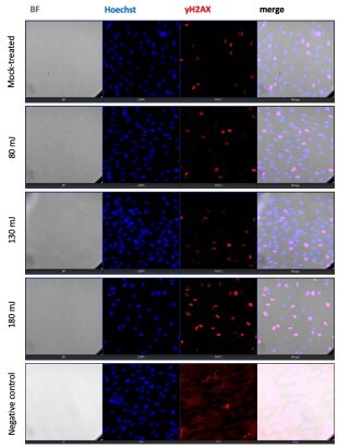
Naświetlanie indukowało zależne od dawki zahamowanie proliferacji, istotne w grupie 180 mJ od 10. godziny, a we wszystkich naświetlanych grupach od 14. godziny (p ≤ 0,03), nasilające się wraz ze wzrostem energii (p < 0,001). Apoptoza i nekroza nie uległy istotnym zmianom przy 80 i 130 mJ (p > 0,05), natomiast 180 mJ spowodowało wyraźny wzrost obu parametrów (p < 0,001). Ekspresja γH2AX pozostawała niezmieniona przy niższych energiach, lecz była istotnie podwyższona przy 180 mJ (p < 0,001).
Wnioski:
Wyniki wskazują na biologiczny punkt przejścia między 130 a 180 mJ, charakteryzujący się zatrzymaniem proliferacji, aktywacją szlaków śmierci komórkowej oraz nasiloną sygnalizacją uszkodzeń DNA związaną z γH2AX. Uzyskane dane mają charakter wskazujący, a nie definitywny, i wymagają walidacji w udoskonalonych modelach eksperymentalnych przed zastosowaniem klinicznym.
Odpowiedź biologiczna fibroblastów na naświetlanie laserem Er:YAG jest zależna od dawki, jednak próg między ekspozycją biologicznie tolerowaną a cytotoksyczną pozostaje niedostatecznie określony. Celem badania jest ocena zależnych od dawki efektów naświetlania laserem Er:YAG (80–180 mJ) na proliferację, apoptozę/nekrozę oraz sygnalizację uszkodzeń DNA związaną z γH2AX w ludzkich fibroblastach w warunkach in vitro.
Materiał i metody:
Ludzkie fibroblasty napletka (human foreskin fibroblasts – HFF-1) poddano naświetlaniu laserem Er:YAG (2940 nm; 10 Hz; 300 µs; 3 min) przy energiach 80, 130 lub 180 mJ i porównano z grupą kontrolną, poddaną procedurze pozorowanej. Proliferację oceniano za pomocą 24-godzinnego mikroskopowego obrazowania poklatkowego z fluorescencją. Apoptozę i nekrozę oznaczano barwieniem Aneksyną V/EthD-III, a uszkodzenia DNA za pomocą immunofluorescencyjnego wykrywania γH2AX. Przeprowadzono trzy niezależne repliki biologiczne; zastosowano analizę ANOVA z testami post hoc (p < 0,05).
Wyniki:
Naświetlanie indukowało zależne od dawki zahamowanie proliferacji, istotne w grupie 180 mJ od 10. godziny, a we wszystkich naświetlanych grupach od 14. godziny (p ≤ 0,03), nasilające się wraz ze wzrostem energii (p < 0,001). Apoptoza i nekroza nie uległy istotnym zmianom przy 80 i 130 mJ (p > 0,05), natomiast 180 mJ spowodowało wyraźny wzrost obu parametrów (p < 0,001). Ekspresja γH2AX pozostawała niezmieniona przy niższych energiach, lecz była istotnie podwyższona przy 180 mJ (p < 0,001).
Wnioski:
Wyniki wskazują na biologiczny punkt przejścia między 130 a 180 mJ, charakteryzujący się zatrzymaniem proliferacji, aktywacją szlaków śmierci komórkowej oraz nasiloną sygnalizacją uszkodzeń DNA związaną z γH2AX. Uzyskane dane mają charakter wskazujący, a nie definitywny, i wymagają walidacji w udoskonalonych modelach eksperymentalnych przed zastosowaniem klinicznym.
FINANSOWANIE
Na badania nie otrzymano żadnego zewnętrznego dofinansowania.
KONFLIKT INTERESÓW
Autorzy deklarują brak konfliktu interesów.
Oświadczenie o wykorzystaniu narzędzi AI: Do korekty językowej wykorzystano narzędzie Grammarly AI.
REFERENCJE (33)
1.
Lin T, Yu CC, Liu CM, Hsieh PL, Liao YW, Yu CH, et al. Er:YAG laser promotes proliferation and wound healing capacity of human periodontal ligament fibroblasts through galectin-7 induction. J Formos Med Assoc. 2021;120(1 Pt 2):388–394. doi: 10.1016/j.jfma.2020.06.005.
2.
Pourzarandian A, Watanabe H, Ruwanpura SM, Aoki A, Noguchi K, Ishikawa I. Er:YAG laser irradiation increases prostaglandin E production via the induction of cyclooxygenase-2 mRNA in human gingival fibroblasts. J Periodontal Res. 2005;40(2):182–186. doi: 10.1111/j.1600-0765.2005.00789.x.
3.
Pourzarandian A, Watanabe H, Ruwanpura SM, Aoki A, Ishikawa I. Effect of low-level Er:YAG laser irradiation on cultured human gingival fibroblasts. J Periodontol. 2005;76(2):187–193. doi: 10.1902/jop.2005.76.2.187.
4.
Hympanova L, Mackova K, El-Domyati M, Vodegel E, Roovers JP, Bosteels J, et al. Effects of non-ablative Er:YAG laser on the skin and the vaginal wall: systematic review of the clinical and experimental literature. Int Urogynecol J. 2020;31(12):2473–2484. doi: 10.1007/s00192-020-04452-9.
5.
Ogita M, Tsuchida S, Aoki A, Satoh M, Kado S, Sawabe M, et al. Increased cell proliferation and differential protein expression induced by low-level Er:YAG laser irradiation in human gingival fibroblasts: proteomic analysis. Lasers Med Sci. 2015;30(7):1855–1866. doi: 10.1007/s10103-014-1691-4.
6.
Zaitsev AE, Asanov ON. Erbium:yttrium aluminium garnet (Er:YAG) laser therapy versus sharp debridement in the management of chronic ulcers of the lower extremity: A randomized controlled trial. Int Wound J. 2025;22(6):e70688. doi: 10.1111/iwj.70688.
7.
Maghfour J, Ozog DM, Mineroff J, Jagdeo J, Kohli I, Lim HW. Photobiomodulation CME part I: Overview and mechanism of action. J Am Acad Dermatol. 2024;91(5):793–802. doi: 10.1016/j.jaad.2023.10.073.
8.
de Freitas LF, Hamblin MR. Proposed mechanisms of photobiomodulation or low-level light therapy. IEEE J Sel Top Quantum Electron. 2016;22(3):7000417. doi: 10.1109/JSTQE.2016.2561201.
9.
Guo Z, Yuan K. The application of light emitting diode (LED) in cosmetic dermatology. Photodermatol Photoimmunol Photomed. 2025;41(5):e70041. doi: 10.1111/phpp.70041.
10.
Dompe C, Moncrieff L, Matys J, Grzech-Leśniak K, Kocherova I, Bryja A, et al. Photobiomodulation-Underlying Mechanism and Clinical Applications. J Clin Med. 2020;9(6):1724. doi: 10.3390/jcm9061724.
11.
Glass GE. Photobiomodulation: the clinical applications of low-level light therapy. Aesthet Surg J. 2021;41(6):723–738. doi: 10.1093/asj/sjab025.
12.
Wang X, Tian F, Soni SS, Gonzalez-Lima F, Liu H. Interplay between up-regulation of cytochrome-c-oxidase and hemoglobin oxygenation induced by near-infrared laser. Sci Rep. 2016;6:30540. doi: 10.1038/srep30540.
13.
Modena DAO, Miranda ACG, Grecco C, Liebano RE, Cordeiro RCT, Guidi RM. Efficacy, safety, and guidelines of application of the fractional ablative laser erbium:YAG 2940 nm and non-ablative laser erbium glass in rejuvenation, skin spots, and acne in different skin phototypes: a systematic review. Lasers Med Sci. 2020;35(9):1877–1888. doi: 10.1007/s10103-020-03046-7.
14.
Pan TL, Wang PW, Lee WR, Fang CL, Chen CC, Huang CM, et al. Systematic evaluations of skin damage irradiated by an erbium:YAG laser: histopathologic analysis, proteomic profiles, and cellular response. J Dermatol Sci. 2010;58(1):8–18. doi: 10.1016/j.jdermsci.2010.02.001.
15.
Weniger FG, Weidman AA, Barrero Castedo CE. Full-field erbium:YAG laser resurfacing: complications and suggested safety parameters. Aesthet Surg J. 2020;40(10):NP374–NP385. doi: 10.1093/asj/sjz319.
16.
Tanzi EL, Alster TS. Side effects and complications of variable-pulsed erbium:yttrium–aluminum–garnet laser skin resurfacing: extended experience with 50 patients. Plast Reconstr Surg. 2003;111(4):1524–1529. doi: 10.1097/01.PRS.0000049647.65948.50.
17.
Alsaad SM, Ross EV, Smith WJ, DeRienzo DP. Analysis of depth of ablation, thermal damage, wound healing, and wound contraction with erbium:YAG laser in a Yorkshire pig model. J Drugs Dermatol. 2015;14(11):1245–1252.
18.
Fiegler-Rudol J, Kępa M, Skaba D, Wiench R. Er:YAG laser energy optimization for reducing single-species microbial growth on agar surfaces in vitro. Pathogens. 2025;14(12):1287. doi: 10.3390/pathogens14121287.
19.
Trakarnphornsombat W, Kimura H. Live-cell tracking of γ-H2AX kinetics reveals the distinct modes of ATM and DNA-PK in the immediate response to DNA damage. J Cell Sci. 2023;136(8):jcs260698. doi: 10.1242/jcs.260698.
20.
Prabhu KS, Kuttikrishnan S, Ahmad N, Habeeba U, Mariyam Z, Suleman M, et al. H2AX: A key player in DNA damage response and a promising target for cancer therapy. Biomed Pharmacother. 2024;175:116663. doi: 10.1016/j.biopha.2024.116663.
21.
Mah LJ, El-Osta A, Karagiannis TC. gammaH2AX: a sensitive molecular marker of DNA damage and repair. Leukemia. 2010; 24(4):679–686. doi: 10.1038/leu.2010.6.
22.
Rahmanian N, Shokrzadeh M, Eskandani M. Recent advances in γH2AX biomarker-based genotoxicity assays: A marker of DNA damage and repair. DNA Repair. 2021;108:103243. doi: 10.1016/j.dnarep.2021.103243.
23.
Kinner A, Wu W, Staudt C, Iliakis G. Gamma-H2AX in recognition and signaling of DNA double-strand breaks in the context of chromatin. Nucleic Acids Res. 2008;36(17):5678–5694. doi: 10.1093/nar/gkn550.
24.
Surova O, Zhivotovsky B. Various modes of cell death induced by DNA damage. Oncogene. 2013;32(33):3789–3797. doi: 10.1038/onc.2012.556.
25.
Matt S, Hofmann TG. The DNA damage-induced cell death response: a roadmap to kill cancer cells. Cell Mol Life Sci. 2016;73(15):2829–2850. doi: 10.1007/s00018-016-2130-4.
26.
Borges HL, Linden R, Wang JY. DNA damage-induced cell death: lessons from the central nervous system. Cell Res. 2008;18(1):17–26. doi: 10.1038/cr.2007.110.
27.
Tu HC, Ren D, Wang GX, Chen DY, Westergard TD, Kim H, et al. The p53-cathepsin axis cooperates with ROS to activate programmed necrotic death upon DNA damage. Proc Natl Acad Sci U S A. 2009;106(4):1093–1098. doi: 10.1073/pnas.0808173106.
28.
Lynnyk A, Lunova M, Jirsa M, Egorova D, Kulikov A, Kubinová Š, et al. Manipulating the mitochondria activity in human hepatic cell line Huh7 by low-power laser irradiation. Biomed Opt Express. 2018;9(3):1283–1300. doi: 10.1364/BOE.9.001283.
29.
Wu S, Xing D, Wang F, Chen T, Chen WR. Mechanistic study of apoptosis induced by high-fluence low-power laser irradiation using fluorescence imaging techniques. J Biomed Opt. 2007;12(6):064015. doi: 10.1117/1.2804923.
30.
Wu S, Xing D, Gao X, Chen WR. High fluence low-power laser irradiation induces mitochondrial permeability transition mediated by reactive oxygen species. J Cell Physiol. 2009;218(3):603–611. doi: 10.1002/jcp.21636.
31.
Lukac M, Zorman A, Lukac N, Perhavec T, Tasic B. Characteristics of non-ablative resurfacing of soft tissues by repetitive Er:YAG laser pulse irradiation. Lasers Surg Med. 2021;53(9):1266–1278. doi: 10.1002/lsm.23402.
32.
Ross EV, McKinlay JR, Sajben FP, Miller CH, Barnette DJ, Meehan KJ, et al. Use of a novel erbium laser in a Yucatan minipig: a study of residual thermal damage, ablation, and wound healing as a function of pulse duration. Lasers Surg Med. 2002;30(2):93–100. doi: 10.1002/lsm.10030.
33.
Chung H, Dai T, Sharma SK, Huang YY, Carroll JD, Hamblin MR. The nuts and bolts of low-level laser (light) therapy. Ann Biomed Eng. 2012;40(2):516–533. doi: 10.1007/s10439-011-0454-7.
Udostępnij
ARTYKUŁ POWIĄZANY
| eISSN: | 1734-025X |

Śląski Uniwersytet Medyczny w Katowicach, jako Operator Serwisu annales.sum.edu.pl, przetwarza dane osobowe zbierane podczas odwiedzania Serwisu. Realizacja funkcji pozyskiwania informacji o Użytkownikach i ich zachowaniu odbywa się poprzez dobrowolnie wprowadzone w formularzach informacje, zapisywanie w urządzeniach końcowych plików cookies (tzw. ciasteczka), a także poprzez gromadzenie logów serwera www, będącego w posiadaniu Operatora Serwisu. Dane, w tym pliki cookies, wykorzystywane są w celu realizacji usług zgodnie z Polityką prywatności.
Możesz wyrazić zgodę na przetwarzanie danych w tych celach, odmówić zgody lub uzyskać dostęp do bardziej szczegółowych informacji.
Możesz wyrazić zgodę na przetwarzanie danych w tych celach, odmówić zgody lub uzyskać dostęp do bardziej szczegółowych informacji.